Otomycosis in Lisbon: ear fungus, symptoms and treatment
Otomycosis is a fungal infection of the external ear canal, also known as fungal otitis externa or, more informally, ear fungus.
It can cause intense itching, a feeling of a blocked ear, discharge, discomfort, pain and reduced hearing. The main agents associated with otomycosis include fungi of the genera Aspergillus and Candida. In general, Aspergillus is identified more frequently, but the distribution of agents varies between regions, populations and clinical series.
English-speaking ENT consultation in Lisbon — Saldanha
Otoscopy and medical assessment · Cleaning or micro-suction when indicated
ENT consultation: €130 — Direct WhatsApp booking in English
What can otomycosis look like?
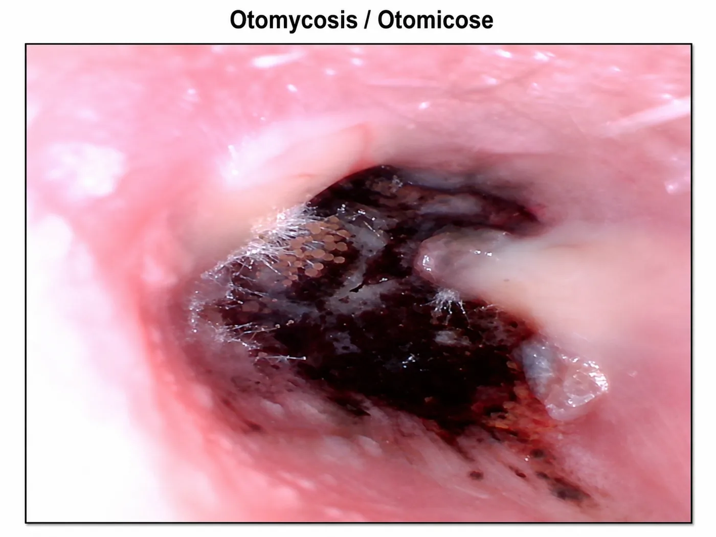
The appearance of otomycosis can vary considerably. Some infections show whitish or cotton-like material; others may show grey, brown or black material.
A dark, granular or filamentous appearance may raise suspicion of filamentous fungi, including Aspergillus species, but the clinical appearance alone does not reliably determine which fungus is responsible.
It is particularly important not to assume that black material necessarily corresponds to Aspergillus niger. The so-called black aspergilli include several species of the Nigri section with similar morphological features.
From the ear to the microscope
On otoscopy we observe the overall appearance of the fungal colony and the material accumulated in the ear canal.
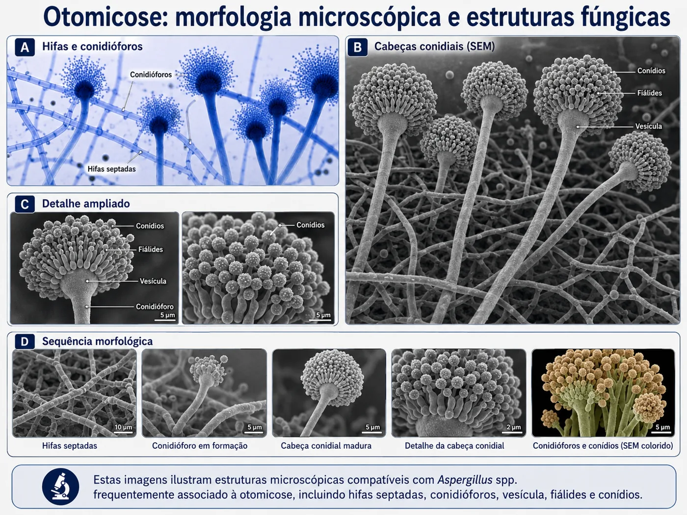
Under the microscope, much smaller structures may be seen, such as hyphae, conidiophores, vesicles, phialides and conidia.
The similarity between certain macroscopic patterns and fungal morphology is clinically interesting and may guide suspicion, but identifying a species requires appropriate microbiological methods when clinically necessary.
“What we see on otoscopy is the fungal colony; the conidiophore is one of the microscopic structures that helps explain its morphology.”
What are the microscopic structures of Aspergillus?
Some of the most frequent causes of otomycosis belong to the genus Aspergillus. Candida species are another relevant group of agents, although they have a very different microscopic morphology.
Aspergillus are filamentous fungi made up of a network of hyphae. During asexual reproduction they can form specialised structures called conidiophores.
The conidiophore usually ends in a swelling called the vesicle. On this, phialides are organised — directly or through metulae, depending on the species — giving rise to chains of small reproductive structures called conidia.
Together these form what is known as the conidial head.
These structures are microscopic. Therefore, although an Aspergillus colony in the ear may appear granular, filamentous or dotted on otoscopy, one should not assume that each visible dot corresponds directly to an individual conidiophore or conidium.
Aspergillus and Candida: different morphologies
Although Aspergillus is one of the most frequently identified genera in otomycosis, Candida species are also an important cause of fungal infection of the ear canal.
The two groups have quite different morphologies under the microscope.
Aspergillus is a filamentous fungus. It forms a network of hyphae and can develop specialised reproductive structures called conidiophores. These end in a vesicle on which phialides are organised — directly or through metulae, depending on the species — responsible for forming the conidia.
Candida predominantly shows a yeast-like morphology. The cells are usually round or oval and reproduce by budding. Some species, particularly Candida albicans, can also form pseudohyphae and true hyphae under certain conditions.
This difference is important: the conidiophores characteristic of Aspergillus are not the typical form of organisation of Candida.
Thus, the term otomycosis encompasses infections caused by fungi with quite different morphologies, capable of producing similar clinical manifestations in the ear canal.
Among the Candida species described in cases of otomycosis are Candida albicans and Candida parapsilosis, among others. The relative frequency of each species varies between studies and populations.
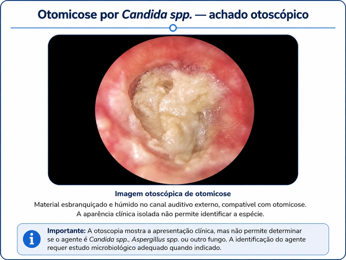
As with Aspergillus, the otoscopic appearance does not allow reliable identification of a Candida species. Microbiological identification, when clinically necessary, requires appropriate study of the collected material.
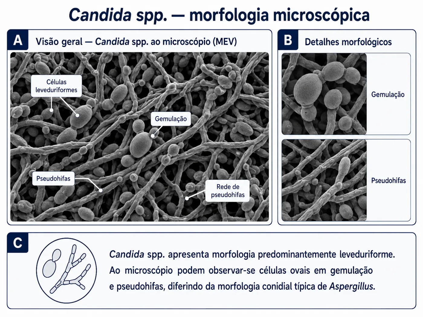
Two different forms of fungus
Aspergillus
Filamentous fungus → hyphae → conidiophore → vesicle → phialides → conidia.
Candida
Predominantly yeast-like fungus → oval/round budding cells → possibility of pseudohyphae and, in some species and conditions, true hyphae.
Both can be involved in otomycosis, but they have a very different microscopic organisation.
Symptoms of ear fungus
The most common symptoms include:
- intense itching inside the ear;
- a feeling of a blocked or full ear;
- reduced or muffled hearing;
- persistent discharge or moisture;
- discomfort or pain;
- a sensation of material inside the ear;
- tinnitus in some cases.
Itching can be particularly marked in fungal infections of the ear canal.
Why does ear fungus appear?
The ear canal has natural protective mechanisms. Some situations upset this balance and can make it easier for fungi to grow.
Associated factors include:
- repeated entry of water into the ear;
- swimming or very humid environments;
- use of cotton buds;
- frequent manipulation of the ear canal;
- minor trauma to the ear skin;
- prolonged or repeated use of certain ear drops;
- hearing aids, earphones or plugs;
- dermatitis or eczema of the canal;
- diabetes or immune changes in certain patients;
- a history of otitis externa.
Ear fungus or simply earwax?
Not all dark material inside the ear is otomycosis.
A blocked ear can result from:
- an earwax plug;
- bacterial otitis externa;
- otomycosis;
- eczema or flaking of the canal skin;
- secretions coming from the middle ear;
- a foreign body;
- other changes of the ear canal or eardrum.
For this reason, using antifungal or antibiotic drops without first examining the ear may not resolve the problem.
Otoscopy allows us to distinguish earwax from inflammatory or fungal material and, at the same time, assess the state of the eardrum. When the problem is mainly wax, see also the page on ear cleaning.
How is the diagnosis made?
The assessment starts with the history of symptoms and direct examination of the ear canal and eardrum.
When there is material in the canal, it may need to be removed progressively through suction or appropriate instruments under direct vision.
In recurrent, treatment-resistant or atypical situations, microbiological testing or culture may be indicated to identify the agent.
How is otomycosis treated?
Treatment is decided after examining the ear canal and the eardrum.
It often includes:
- careful cleaning of the ear canal, removing fungal material and secretions;
- keeping the ear dry;
- topical antifungal treatment when indicated;
- control of predisposing factors;
- reassessment when there is persistence or recurrence.
The choice of drops should not be made by visual appearance or trial alone, because it is important to know whether the eardrum is intact and whether there is any associated condition.
What not to do if you suspect otomycosis
Avoid introducing into the ear:
- cotton buds;
- tweezers or other objects;
- alcohol, oils or home remedies;
- antibiotic or antifungal drops chosen without medical assessment;
- water to try to wash out the fungus.
Manipulation can traumatise the skin, push material deeper and perpetuate inflammation.
Can otomycosis come back?
It can.
Recurrence is possible, especially when predisposing factors remain, there is a lot of moisture in the canal or fungal material persists.
For this reason, some patients may need more than one visit until the canal is adequately clean and the skin has recovered.
Warning signs — when to seek urgent care
- severe or progressive pain;
- fever or significant deterioration in general condition;
- sudden hearing loss;
- intense vertigo;
- abundant blood or discharge;
- significant swelling of the ear or surrounding area;
- facial weakness;
- diabetes or immunosuppression associated with severe pain or an unfavourable course.
These signs do not confirm a diagnosis, but warrant prompt medical assessment. In an emergency, call 112.
How the ENT consultation works
During the consultation, the ear canal and eardrum are examined with otoscopy and, when necessary, microscopy.
When there is fungal material or secretions, they can be carefully removed by suction or instruments, allowing the eardrum to be seen afterwards and the most appropriate treatment to be defined.
If there is suspicion of hearing loss that warrants further investigation, complementary hearing tests are referred to an appropriate centre.
Consultation for otomycosis in Lisbon
Dr. Pedro Stapleton-Garcia — ENT specialist
Private practice in Saldanha, Lisbon. Avenida 5 de Outubro, 16, 2nd floor, right. About a 5-minute walk from Saldanha metro station.
ENT consultation: €130
Consultations in English, Portuguese, French, Spanish and Czech.
Frequently asked questions about otomycosis
How do I know if I have ear fungus?
Intense itching, a blocked ear, discharge and whitish, greyish or dark material may suggest otomycosis. However, earwax, eczema and bacterial infections can cause similar symptoms. Otoscopic examination is important to tell them apart.
Are otomycosis and fungal otitis the same thing?
Yes. Otomycosis is the medical term for a fungal infection of the external ear canal and is often called fungal otitis externa.
Can Aspergillus cause ear fungus?
Yes. Aspergillus species are among the main fungi associated with otomycosis. Candida and other fungi can also be involved.
Does black material in the ear mean Aspergillus niger?
Not necessarily. Black or granular material may raise suspicion of filamentous fungi and some Aspergillus species can show dark pigmentation. However, several morphologically similar species exist and the otoscopic appearance cannot reliably identify Aspergillus niger or any other species.
What is a conidiophore?
It is a specialised microscopic structure used by certain filamentous fungi to produce conidia. In Aspergillus, the conidiophore ends in a vesicle on which phialides and, in certain species, metulae are organised.
Is it necessary to clean the ear?
Often yes. Removing fungal material, debris and secretions improves contact of the local treatment with the ear canal and allows the eardrum to be seen properly.
Can I treat otomycosis at home?
It is not advisable to start drops or home remedies before knowing whether otomycosis is really present and whether the eardrum is intact.
Can ear fungus cause hearing loss?
The build-up of material in the canal can partially block the passage of sound and cause a temporary conductive hearing loss. If there is sudden or disproportionate hearing loss, medical assessment is needed.
Can Candida cause otomycosis?
Yes. Candida species can cause fungal infections of the ear canal and, together with Aspergillus, are among the fungi most frequently associated with otomycosis. Described species include Candida albicans and Candida parapsilosis, although the distribution varies between populations.
Does Candida have conidiophores like Aspergillus?
No. The conidiophores with vesicle, phialides and conidia described on this page are characteristic of the morphology of Aspergillus. Candida predominantly shows yeast-like cells that reproduce by budding and can form pseudohyphae and, under certain conditions, true hyphae.
Related pages
You may also be interested in: ear cleaning · English-speaking ENT in Lisbon · book an appointment · Versão portuguesa
Medical content by Dr. Pedro Stapleton-Garcia, ENT specialist, Lisbon.
Last updated: October 2026.
This content is for information purposes only and does not replace an individual medical assessment.